- Retinopathy of prematurity (ROP) occurs as a result of abnormal growth of the blood vessels of the eye, often caused by fluctuations in oxygen levels in the developing retina.
- All premature infants responding to the selection protocol defined by a birth weight of less than 1500 grams and a gestational age of less than 30 weeks are routinely screened for retinopathy of prematurity.
- Depending on the amount of blood vessels that develop abnormally, a classification of the baby’s disease will be made and other tests will be done weekly or every two weeks.
Retinopathy of the premature (ROP) occurs as a result of abnormal growth of the blood vessels of the eye. A premature baby with a birth weight of less than 1,500 grams and a gestational age of less than 30 weeks will be regularly screened to see if he has retinopathy of prematurity.
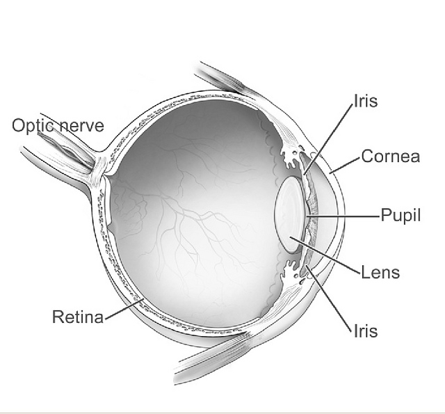
It lines the inside of the eye. At around 16 weeks of pregnancy, the retina begins to develop blood vessels. These usually develop in areas where oxygenation is high, where the vessels already exist, to areas where oxygenation is low, where the vessels are not yet formed. Thus, the retina covers blood vessels gradually and uniformly.
The change in oxygen content in the developing retina is an important factor in the regulation of blood vessel growth. In addition, other complications that interfere with the regular flow of oxygenated blood that the baby may have may also be involved in the normal growth and development of the blood vessels. Retinopathy of premature infants (ROP, also known as retrocrystal fibroplasia or retrolental fibroplasia) is the case in which blood vessels develop abnormally.
How is ROP diagnosed?
All preterm infants responding to the selection protocol defined by a birth weight of less than 1500 grams and a gestational age of less than 30 weeks are routinely screened for ROP. These premature babies will probably be examined for the first time four to six weeks after birth. The ophthalmologist will use scours to dilate the pupil, which will allow a better view of the inside of the eye.
Depending on the amount of blood vessels that develop abnormally, a classification of the baby’s disease will be made and other tests will be done every one to two weeks depending on various factors. These factors include the severity and position of ROP in the eye, and the rate of progression of blood vessel formation, called vascularization. In the majority of cases, even when retinopathy of prematurity develops, it disappears spontaneously with little effect on vision. However, a small percentage of babies controlled for ROP, about 10%, will develop the condition to such an extent that waiting for it to go away spontaneously is a danger. So we offer these babies a treatment that aims to reverse the progression of ROP.
Stages of the ROP
The severity of ROP is assessed according to the affected area of the retina, also known as the area of vascularization, and the stages of the disease. The lower the number in the area, the larger the area affected by the retina. The stages, or progression of growth of abnormal blood vessels, are defined below.
Stage 1 of the ROP
At stage 1 of the ROP, there is a small gap between the area where there are blood vessels and the one where the blood vessels are not yet formed. At this point, it is possible for the vessels to develop on their own, but the disease must be monitored.
Stage 2 of the ROP
In Stage 2, the space between the regions where there are and where there are no vessels expands into a dividing line. At this stage, the disease can still disappear, however, it can also develop in stage 3.
Stage 3 of the ROP
At stage 3, new blood vessels begin to develop along the dividing line and spread into the clear gel that fills the eye, called the vitreous body. These blood vessels can bleed and form scar tissue.
Stage 4A of the ROP
In stage 4, abnormal blood vessels and scar tissue pull on the retina, partially detaching it. At stage 4A, the center of vision, called fovea, is not affected.
Stage 4B ROP
At stage 4B, the retina is still only partially detached, but the fovea is affected, usually decreasing peripheral vision and central vision to a certain extent.
Stage 5 of the ROP
In stage 5, the retina is completely detached and vision is severely affected.
